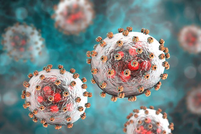
Lassa fever is a viral illness that is far too common in West Africa. Although it can have a mortality rate of 15 percent in severe cases, up to 90% in pregnant women, and causes deafness in a quarter of survivors, there is no vaccine or antiviral to protect against Lassa virus. To save lives, scientists at La Jolla Institute for Immunology (LJI) and Scripps Research are working to understand exactly how Lassa virus replicates within human hosts.
In a new study, published in Proceedings of the National Academy of Sciences, the researchers show how a critical Lassa virus protein, called polymerase, drives infection by harnessing a cellular protein in human hosts. Their work suggests future therapies could target this interaction to treat patients.
“There is no antiviral drug that specifically targets Lassa virus,” explains study first author Jingru Fang, a joint LJI and Scripps Research graduate student. “That’s why it’s important for researchers to identify potential druggable targets on this virus to combat infection.”
Lassa virus encodes only four viral proteins. One of them, the polymerase, directs the process of virus genome replication and gene expression to produce the materials the virus needs to spread to new host cells. If one can stop virus polymerase, one can stop infection.
Together with study senior authors LJI President and CEO Erica Ollmann Saphire, Ph.D., and Scripps Research Professor Juan De La Torre, Ph.D., Fang led the hunt for host cellular proteins that may act as Lassa polymeras’s partners in crime.
The hunt for Lassa’s helper
Fang and her colleagues engineered Lassa virus polymerase to carry an enzymatic tag that labels polymerase-interacting host proteins with a special chemical handle. The researchers then fished out host proteins with this chemical handle and used a technique called mass spectrometry to identify these host proteins that interact with Lassa virus polymerase.
“It is like defining the Lassa virus polymerase social network, which allows you to go after partners,” says De La Torre.
In collaboration with Professor Alexander Bukreyev, Ph.D., and colleagues at University of Texas Medical Branch (UTMB), the team carried out a “function screen” using live Lassa virus. This work, carried out inside a high-containment laboratory, revealed which of these host proteins could be important for Lassa infection. Among a total of 42 host proteins that interact with Lassa polymerase, the team focused on one druggable target: GSPT1. The team showed that GSPT1 is physically and functionally linked to Lassa virus polymerase and can facilitate Lassa virus infection.
This study is the first to uncover molecular cross-talks between Lassa virus polymerase and cellular proteins. However, it is the second-ever time the host protein GSPT1 has been linked to virus infection. The first was a recent Cell Reports study showing viral polymerase hijacking GSPT1 in Ebola virus infections—research also led by Saphire, De La Torre, and Fang.
“If we could find a way to either disrupt the link between GSPT1 and Lassa polymerase, or if we could simply remove GSPT1 protein, we could stop Lassa virus infection,” says Fang.
Eyeing a new drug for Lassa
To their surprise, the team found a drug candidate, called CC-90009, which has been shown to destroy GSPT1 proteins and is currently being examined as a cancer therapy in clinical trials.
To see whether they can repurpose the existing GSPT1 inhibitor against Lassa infection, Research Associate Colette Pietzsch, from Bukreyev group at UTMB, added CC-90009 to Lassa-infected human liver cells inside a high-containment laboratory. This experiment showed that CC-90009 treatment significantly reduced Lassa virus growth without obvious cell toxicity.
The researchers say it is feasible that this same small molecule drug could double as an Ebola virus therapy, and their Cell Reports data suggest that CC-90009 can reduce virus titer at later time points of Ebola virus infection.
“Translating this finding to therapeutic interventions will still take time,” says Fang. “We need to confirm that CC-90009 can inhibit Lassa and Ebola virus replication in animal models of infection, but we at least we have a starting point.”
Table of Contents
Key facts
- Lassa fever is an acute viral haemorrhagic illness of 2-21 days duration that occurs in West Africa.
- The Lassa virus is transmitted to humans via contact with food or household items contaminated with rodent urine or faeces.
- Person-to-person infections and laboratory transmission can also occur, particularly in hospitals lacking adequate infection prevention and control measures.
- Lassa fever is known to be endemic in Benin, Ghana, Guinea, Liberia, Mali, Sierra Leone, and Nigeria, but probably exists in other West African countries as well.
- The overall case-fatality rate is 1%. Observed case-fatality rate among patients hospitalized with severe cases of Lassa fever is 15%.
- Early supportive care with rehydration and symptomatic treatment improves survival.
Background
Though first described in the 1950s, the virus causing Lassa disease was not identified until 1969. The virus is a single-stranded RNA virus belonging to the virus family Arenaviridae.
About 80% of people who become infected with Lassa virus have no symptoms. 1 in 5 infections result in severe disease, where the virus affects several organs such as the liver, spleen and kidneys.
Lassa fever is a zoonotic disease, meaning that humans become infected from contact with infected animals. The animal reservoir, or host, of Lassa virus is a rodent of the genus Mastomys, commonly known as the “multimammate rat.” Mastomys rats infected with Lassa virus do not become ill, but they can shed the virus in their urine and faeces.
Because the clinical course of the disease is so variable, detection of the disease in affected patients has been difficult. When presence of the disease is confirmed in a community, however, prompt isolation of affected patients, good infection prevention and control practices, and rigorous contact tracing can stop outbreaks.
Lassa fever is known to be endemic in Benin (where it was diagnosed for the first time in November 2014), Ghana (diagnosed for the first time in October 2011), Guinea, Liberia, Mali (diagnosed for the first time in February 2009), Sierra Leone, and Nigeria, but probably exists in other West African countries as well.
Symptoms of Lassa fever
The incubation period of Lassa fever ranges from 6–21 days. The onset of the disease, when it is symptomatic, is usually gradual, starting with fever, general weakness, and malaise. After a few days, headache, sore throat, muscle pain, chest pain, nausea, vomiting, diarrhoea, cough, and abdominal pain may follow. In severe cases facial swelling, fluid in the lung cavity, bleeding from the mouth, nose, vagina or gastrointestinal tract and low blood pressure may develop.
Protein may be noted in the urine. Shock, seizures, tremor, disorientation, and coma may be seen in the later stages. Deafness occurs in 25% of patients who survive the disease. In half of these cases, hearing returns partially after 1–3 months. Transient hair loss and gait disturbance may occur during recovery.
Death usually occurs within 14 days of onset in fatal cases. The disease is especially severe late in pregnancy, with maternal death and/or fetal loss occurring in more than 80% of cases during the third trimester.
Transmission
Humans usually become infected with Lassa virus from exposure to urine or faeces of infected Mastomys rats. Lassa virus may also be spread between humans through direct contact with the blood, urine, faeces, or other bodily secretions of a person infected with Lassa fever. There is no epidemiological evidence supporting airborne spread between humans. Person-to-person transmission occurs in both community and health-care settings, where the virus may be spread by contaminated medical equipment, such as re-used needles. Sexual transmission of Lassa virus has been reported.
Lassa fever occurs in all age groups and both sexes. Persons at greatest risk are those living in rural areas where Mastomys are usually found, especially in communities with poor sanitation or crowded living conditions. Health workers are at risk if caring for Lassa fever patients in the absence of proper barrier nursing and infection prevention and control practices.
Diagnosis
Because the symptoms of Lassa fever are so varied and non-specific, clinical diagnosis is often difficult, especially early in the course of the disease. Lassa fever is difficult to distinguish from other viral haemorrhagic fevers such as Ebola virus disease as well as other diseases that cause fever, including malaria, shigellosis, typhoid fever and yellow fever.
Definitive diagnosis requires testing that is available only in reference laboratories. Laboratory specimens may be hazardous and must be handled with extreme care. Lassa virus infections can only be diagnosed definitively in the laboratory using the following tests:
- reverse transcriptase polymerase chain reaction (RT-PCR) assay
- antibody enzyme-linked immunosorbent assay (ELISA)
- antigen detection tests
- virus isolation by cell culture.
Treatment and prophylaxis
The antiviral drug ribavirin seems to be an effective treatment for Lassa fever if given early on in the course of clinical illness. There is no evidence to support the role of ribavirin as post-exposure prophylactic treatment for Lassa fever.
There is currently no vaccine that protects against Lassa fever.
Prevention and control
Prevention of Lassa fever relies on promoting good “community hygiene” to discourage rodents from entering homes. Effective measures include storing grain and other foodstuffs in rodent-proof containers, disposing of garbage far from the home, maintaining clean households and keeping cats. Because Mastomys are so abundant in endemic areas, it is not possible to completely eliminate them from the environment. Family members should always be careful to avoid contact with blood and body fluids while caring for sick persons.
In health-care settings, staff should always apply standard infection prevention and control precautions when caring for patients, regardless of their presumed diagnosis. These include basic hand hygiene, respiratory hygiene, use of personal protective equipment (to block splashes or other contact with infected materials), safe injection practices and safe burial practices.
Health-care workers caring for patients with suspected or confirmed Lassa fever should apply extra infection control measures to prevent contact with the patient’s blood and body fluids and contaminated surfaces or materials such as clothing and bedding. When in close contact (within 1 metre) of patients with Lassa fever, health-care workers should wear face protection (a face shield or a medical mask and goggles), a clean, non-sterile long-sleeved gown, and gloves (sterile gloves for some procedures).
Laboratory workers are also at risk. Samples taken from humans and animals for investigation of Lassa virus infection should be handled by trained staff and processed in suitably equipped laboratories under maximum biological containment conditions.
On rare occasions, travellers from areas where Lassa fever is endemic export the disease to other countries. Although malaria, typhoid fever, and many other tropical infections are much more common, the diagnosis of Lassa fever should be considered in febrile patients returning from West Africa, especially if they have had exposures in rural areas or hospitals in countries where Lassa fever is known to be endemic. Health-care workers seeing a patient suspected to have Lassa fever should immediately contact local and national experts for advice and to arrange for laboratory testing.
WHO Response
The Ministries of Health of Guinea, Liberia and Sierra Leone, WHO, the Office of United States Foreign Disaster Assistance, the United Nations, and other partners have worked together to establish the Mano River Union Lassa Fever Network. The programme supports these 3 countries in developing national prevention strategies and enhancing laboratory diagnostics for Lassa fever and other dangerous diseases. Training in laboratory diagnosis, clinical management, and environmental control is also included.

